Salivary Gland Removal Surgery Specialist in Sydney
What are the salivary glands, and where are they located?
The salivary glands produce saliva through a system of ducts connected to the oral cavity. There are three paired major salivary glands (parotid, submandibular, sublingual) and hundreds of minor salivary glands which line the oral cavity. The parotid glands are located on either side of face in front of both ears and extend into the upper neck. The submandibular glands are located on both sides underneath the jaw bone. The sublingual glands are located underneath each side of the tongue.
What are the causes of a swelling or lump in the salivary gland?
There are many causes of a swelling or lump in salivary gland. These include:
- salivary stones
- infections – viral and bacterial
- tumours – benign and cancerous
- inflammatory and autoimmune causes – IgG4 disease, sarcoidosis, Sjogren’s syndrome, diabetes
What will happen during my consultation for my salivary problem?
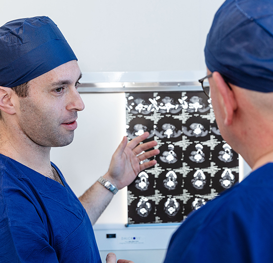
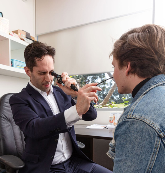
Your consultation will begin with a careful history and physical examination, followed by review of your results including blood tests and scans. A bedside office ultrasound will be conducted to provide real-time diagnostic information. A nasendoscopy (thin fibre-optic camera procedure via the nostril) may be performed under local anaesthesia to examine the back of your throat for related conditions. Further investigations including additional scans and biopsies may be recommended. Treatment options will then be discussed and you will have the opportunity to ask questions in order to make the best decisions for your care.
Do salivary lumps need to be surgically removed?
This depends on the appearance and characteristics of the lump on examination and ultrasound, as well as the results of a biopsy in some patients. Salivary swellings from infection or inflammatory causes can usually be treated with observation or medication and generally do not require surgery. Salivary growths or tumours may require excision if causing concern or symptoms, and sometimes to rule out cancer. Your individual circumstances and need for surgery will be discussed in detail during the consultation.
What is a pleomorphic adenoma?
A pleomorphic adenoma is the most common type of benign salivary tumour, and are most commonly located in the superficial parotid gland. Although benign, the usual recommendation is excision as they have malignant potential with an approximately 10% risk of transformation to cancer over time. Surgery is also warranted to prevent ongoing growth and to confirm the lump is indeed a pleomorphic adenoma and nothing more serious. It is important to remove a margin of normal salivary tissue around the pleomorphic adenoma to prevent it from coming back.
What is a Warthin’s tumour?
Warthin’s tumours are benign tumours which are usually located in the bottom of the parotid gland. They are the second most common benign salivary tumour and can be present in both sides in 20% of cases. Most patients are older, males, and there is an association with smoking. Unlike pleomorphic adenoma, the risk of malignant transformation in Warthin’s tumours is considered highly unlikely and therefore often simple observation is advised rather than surgery. Sometimes surgery is recommended if the tumour is large, growing or causing other symptoms or concerns.
What is the risk of a salivary lump being cancerous?
The risk of salivary lump being cancerous is low for most patients. The risk of cancer depends on the features on history and examination, the appearance on ultrasound and other scans, as well as the results of a tissue biopsy in some patients. Salivary glands contain lymph nodes, and occasionally some cancers, particularly skin cancers, can spread to lymph nodes in the salivary gland, and present as a salivary lump. Your individual results and circumstances will be carefully considered during the consultation to assess the risk of cancer.
What are salivary stones, and how are they treated?
Salivary stones are masses of crystallised minerals that form in the ducts of the salivary gland. The condition is also known as sialolithiasis. The reason why these stones form are unknown but there are some risk factors including taking certain medications (e.g. antidepressants/antihistamines) that reduce saliva production, and dehydration. For many patients, salivary stones can be treated at home with simple measures, but some do require surgery. Surgical intervention usually involves extraction of the stone(s) from the salivary duct via the oral cavity, but in severe cases, complete removal of the salivary gland may be required.
How is parotidectomy performed?
The operation is performed under a general anaesthetic, and nerve monitoring is positioned to aid facial nerve identification and protection. Local anaesthesia is injected and an incision is made in front of the ear and extended behind the ear, and then onto the upper part of the neck. The parotid gland is then carefully exposed and the tumour is removed with a margin of normal salivary tissue if feasible. The most important part of the operation is finding and protecting the facial nerve which passes through the parotid gland. The facial nerve controls the muscles of facial expression and is important for eye closure and lip and mouth movement. The facial nerve is identified and protected with the assistance of intraoperative nerve monitoring. Reconstruction techniques with muscle or fat grafts may be used to fill the cosmetic defect of the excised parotid gland. A drain is usually placed at the end of surgery, and most patients are discharged home the following day. The length of operation varies but usually takes 1-2 hours. Most of the incision is ‘hidden’ in the front ear crease and will not be obviously visible in the medium to long term.
Are the possible complications from parotidectomy?
Most patients undergoing parotidectomy do not have problems. Risks of parotidectomy include general anaesthetic (cardiac and respiratory problems), bleeding, infection, wound problems, salivary leakage, Frey’s syndrome (cheek perspiration when eating) and a small risk of facial nerve weakness. When facial nerve weakness occurs, this is usually temporary and short-lived. The risk of permanent facial nerve weakness is very low and less than 5%. The individual risks vary depending on the nature of the specific case, and will be discussed in more detail during the consultation.
How is submandibular gland surgery performed?
The technique for submandibular surgery depends on the reason for the operation. The surgery is performed under a general anaesthetic and an incision is made approximately 4cm below the jaw bone, after infiltration with a local anaesthetic. The submandibular gland is removed, sometimes with surrounding lymph nodes, and the submandibular duct is tied off. The important nerves nearby the gland are identified and protected – these include the marginal mandibular, the lingual and hypoglossal nerves. A drain is usually placed at the end of surgery, and most patients are discharged home the following day. The length of operation varies but usually takes an hour.
Are the possible complications from submandibular gland surgery?
Most patients undergoing submandibular gland surgery do not have problems. Risks include general anaesthetic (cardiac and respiratory problems), bleeding, infection, wound problems, salivary leakage and a small risk of nerve weakness in the marginal mandibular (facial weakness), lingual (tongue sensation) and hypoglossal (tongue movement) nerves. The risk of these nerve problems are low and usually temporary. The individual risks vary depending on the nature of the specific case, and will be discussed in more detail during the consultation.
What is the recovery like after salivary surgery?
Recovery after salivary surgery is usually straightforward. Most patients have little pain, eat and drink normally and return to work within 7-10 days. Light duties are preferable for 2 weeks after surgery. Further advice regarding recovery in your specific circumstances will be discussed during the preoperative consultation.